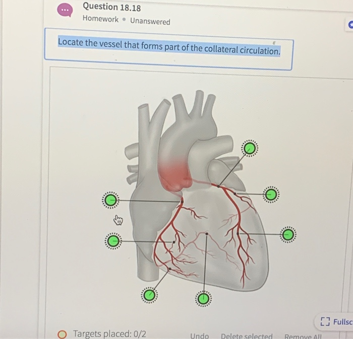
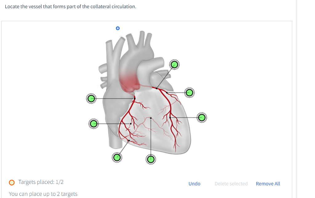
Locate the Vessel That Forms Part of the Collateral Circulation.
Collateral circulation -two vessels interconnect to supply the same area -help supply blood to at risk tissue -very small interconnections are normally found among microscopic branches of arteries referred pain angina. Collateral circulation is the alternate circulation around a blocked artery or vein via another path such as nearby minor vessels.

Pedi Cardiology Anatomy Coronary Veins Coronary Arteries Coronary Arteries Coronary Arteries
When the obstruction is extrahepatic the collateral circulation usually develops toward the portal vein beyond the site of obstruction and thus drains toward the liver hepatopetal collateral circulation.
. As detailed in the anatomy section the azygos vein delivers venous return to the inferior SVC and is the only major vein to feed into the SVC apart from the right and left brachiocephalic veins. This is used by cardiologists to perform a heart bypass when the hearts own. 12 Undo Delete selected.
Eric Stolze Date. Its formation may be. 38 High-quality angiography ideally obtained during breath hold and without panning allowing complete opacification of collateral vessels and obtained in optimal angiographic projections should therefore be encouraged as part of.
The modified vessels through which such circulation occurs. At the apex of the heart where the coronary arteries form. Locate the vessel that forms part of the collateral circulation.
Save or salvage muscular tissue is still lacking. Define arteries arterioles veins venules capillaries a. Collaterals do not form de-novo in the.
However in cirrhosis hepatopetal pathways can be present and in EHPVO hepatofugal pathways can be found. When the coronary arteries narrow to the point that blood flow to the heart muscle is limited coronary artery disease collateral vessels may enlarge and become active. 2 the channels of communication between the blood vessels supplying the heart.
Circulation of blood established through enlargement of minor vessels and anastomosis of vessels with those of adjacent parts when a major vein or artery is functionally impaired as by obstruction also. Order of appearance of collateral. These extra vessels are called collateral blood vessels.
12 Undo Delete selected Remove All You can place up to 2 targets. 02 Undo Delete selected Remove All. Pulmonary arteries are the vessels that transfer blood.
Fullsc O Targets placed. For acute ischemic stroke patients with large vessel occlusion LVO the use of CT and magnetic resonance perfusion imaging to define the ischemic core and penumbra has overshadowed the importance of evaluating the cerebral collateral circulation1 In part this reflects the relative ease with which perfusion imaging can now be interpreted particularly with. Collateral vessels are abnormal blood vessels that connect the aorta with the pulmonary arteries.
Normoxic and ischemic region and that it is. The gold standard involves intracoronary pressure measurements. Unanswered Locate the vessel that forms part of the collateral circulation.
3 major collateral routes include. Collateral vessels are extra blood vessels that connect portions of the same artery or link two different arteries. The new mediacal dictionary.
For example there is a major collateral vessel in the thigh. 1 The other side of the circle of Willis 2 The posterior cerebral circulation 3 The external carotid artery branches Only 50 of patients have a complete circle of Willis due to variants-these patients will not benefit from the. During a stroke leptomeningeal collateral vessels allow limited blood flow when other larger blood vessels provide inadequate blood supply to a.
Collateral circulation is a network of tiny blood vessels and under normal conditions not open. 02 Undo Delete selected Remove All. Happens in one area but has symptom in another place such as a heart attack but hurting in the arm or leg CAD.
The leptomeningeal collateral circulation is a network of small blood vessels in the brain that connects branches of the middle anterior and posterior cerebral arteries with variation in its precise anatomy between individuals. The collateral circulation can be assessed by different methods. In the top panel 3.
Collateral circulation is present in most tissues and provides protection against ischemic injury caused by ischemic stroke coronary atherosclerosis peripheral artery disease and other conditions and diseases 1. It may occur via preexisting vascular redundancy as in the circle of Willis in the brain or it may occur via new branches formed between adjacent blood vessels as in the eye after a retinal embolism or in the brain when moyamoya occurs. Evaluation of the collateral circulation is critical for determining the feasibility of the retrograde approach.
Anatomy and Physiology questions and answers. Three patterns of collateral vessel formation are observed in SVC obstruction all resulting from the location of the blockage relative to the azygos vein 9 10. The aorta is a blood vessel that carries blood from the heart to arteries throughout the body.
March 29 2022 A diagram showing the composition of a blood vessel. Medical Definition of collateral circulation. Is the alternate circulation around a blocked artery or vein via another path such as nearby minor vessels.
Arteries carry blood away from the heart to the tissues. Unanswered Locate the vessel that forms part of the collateral circulation. Locate the vessel that forms part of the collateral circulation.
Flow and pressure can be measured and the control and collateral vessels provide adequate amounts of tissue for many biochemical analyses. Collateral circulation 1 an alternative route provided for the blood by secondary vessels when a primary vessel becomes blocked. Fullsc O Targets placed.
Collateral circulation is a network of alternate circulation around a blocked artery or vein via another path such as nearby minor vessels. Connected to an arterial pressure source and is able to. These alternate blood circulation routes develop in most people and are usually closed to the flow of blood.
The collateral flow velocity is then compared to the flow velocity through the open coronary artery and indicates the percentage of normal blood flow that can be preserved via the collateral circulation in case of an abrupt vessel occlusion. The relevance of these collateral arteries is a matter of ongoing debate but increasing evidence indicates a relevant protective role in patients with coronary artery disease. The answer is yesthere are no collateral circulation in testicle areas the only circulation goes to there is the 5th outlet of oxygenated blood from descending aortait might involved liver on.
Solved Locate The Vessel That Forms Part Of The Collateral Chegg Com
What Is A Collateral Circulation In The Upper Extremity Quora
Solved Question 18 18 Homework Unanswered Locate The Vessel Chegg Com
No comments for "Locate the Vessel That Forms Part of the Collateral Circulation."
Post a Comment